
Medical systems
Smart Assistant Surgeon
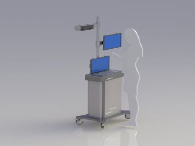
Assists the surgeon during surgery in the operating room. The surgeon can access medical images without touching the mouse or keyboard with voice and motion commands. These images are displayed in two and three dimensions.
DTI && FMRI
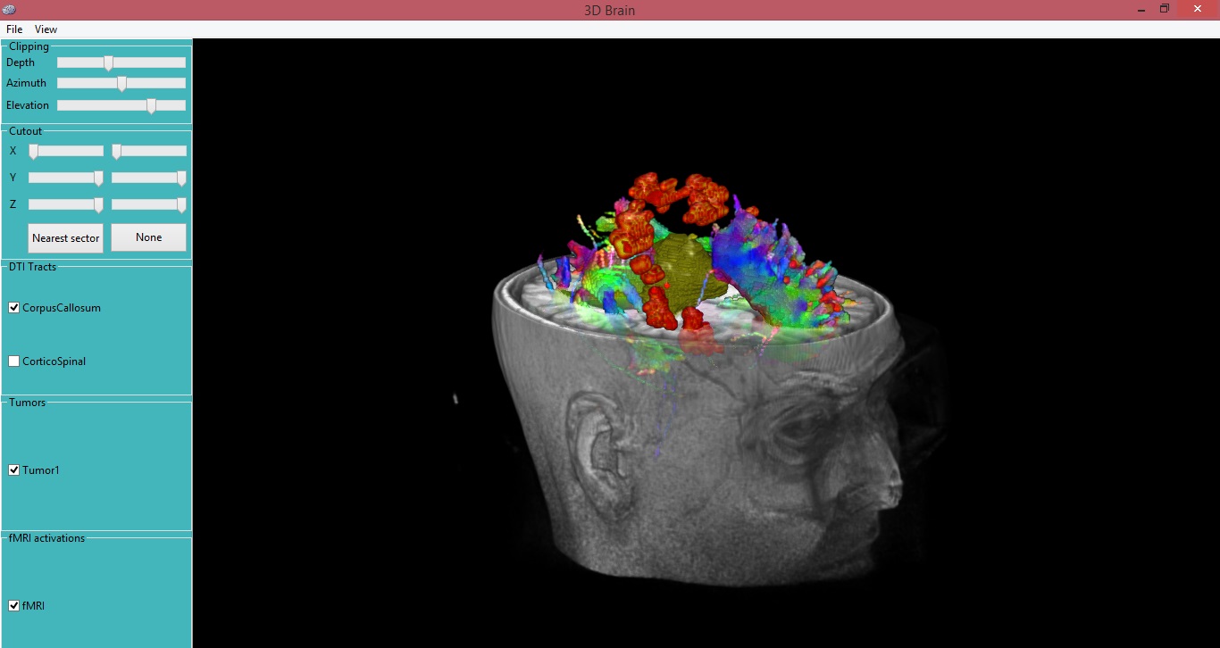
Through this software, the doctor separates the cells and nerve fibers before the surgery in the treatment design stage. To perform surgery during the operation without the slightest damage to the brain.
DAR4DCT
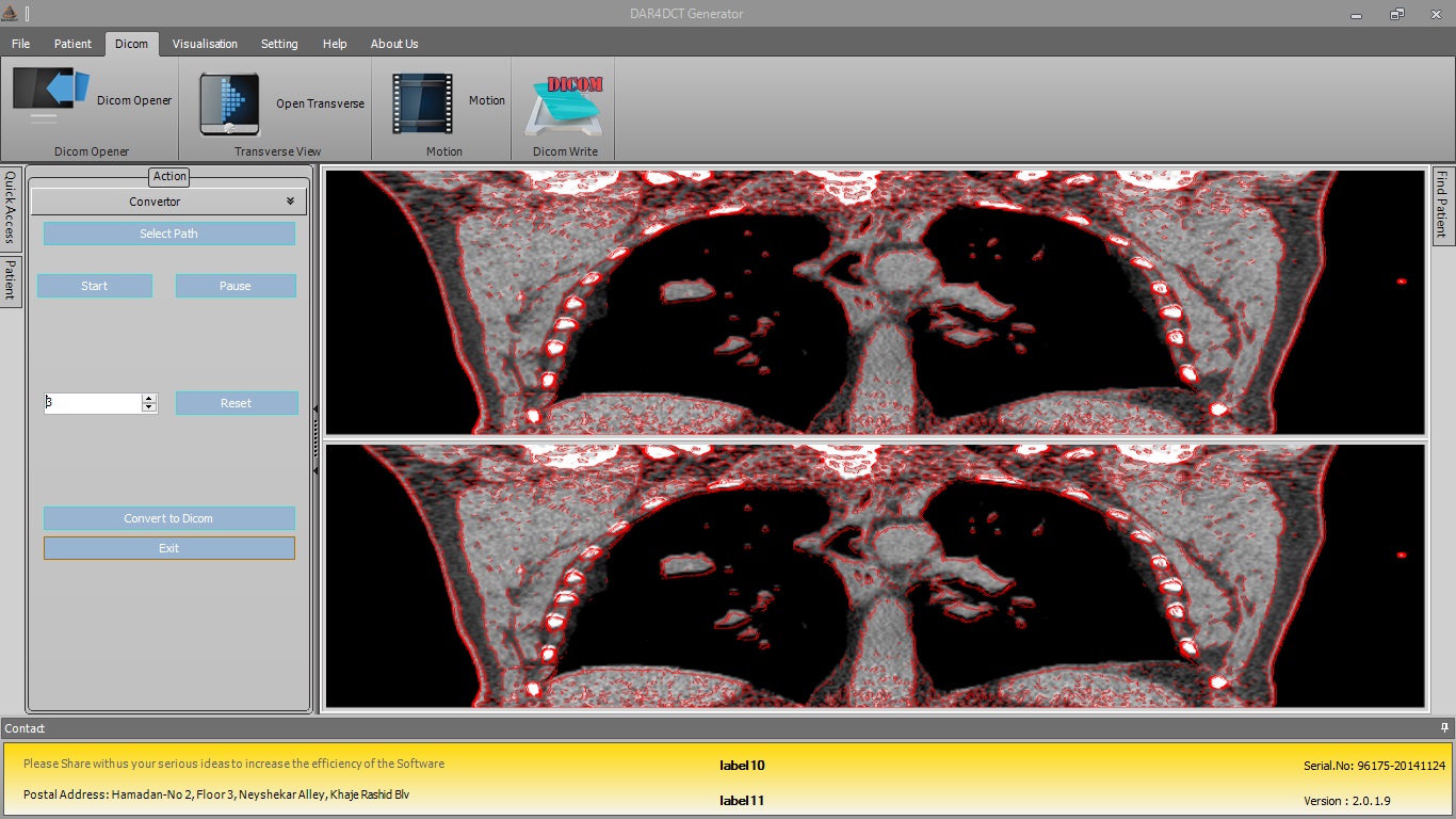
The final product assists the physicist in the treatment design phase. The software simulates the amount of tumor movement in the lungs during inhalation and exhalation.